Diaphragm weakness in the ICU: What do we know?
ICM ARTICLE REVIEW
Diaphragm weakness is common in intensive care. It is associated with increased mortality and failure to wean from ventilation. Its cause in intensive care is often multi-factorial, due to both disease processes and treatment side effects. Currently there are a number of effective preventative strategies and treatments with potential future treatments under investigation.
Aim
This article seeks to review diaphragm weakness in intensive care, aiming to increase awareness of its high prevalence, complex multi-factorial causes and significant consequences as well as the utility of bedside techniques to aid in diagnosis and the efficacy of current and developing treatments.
Key points
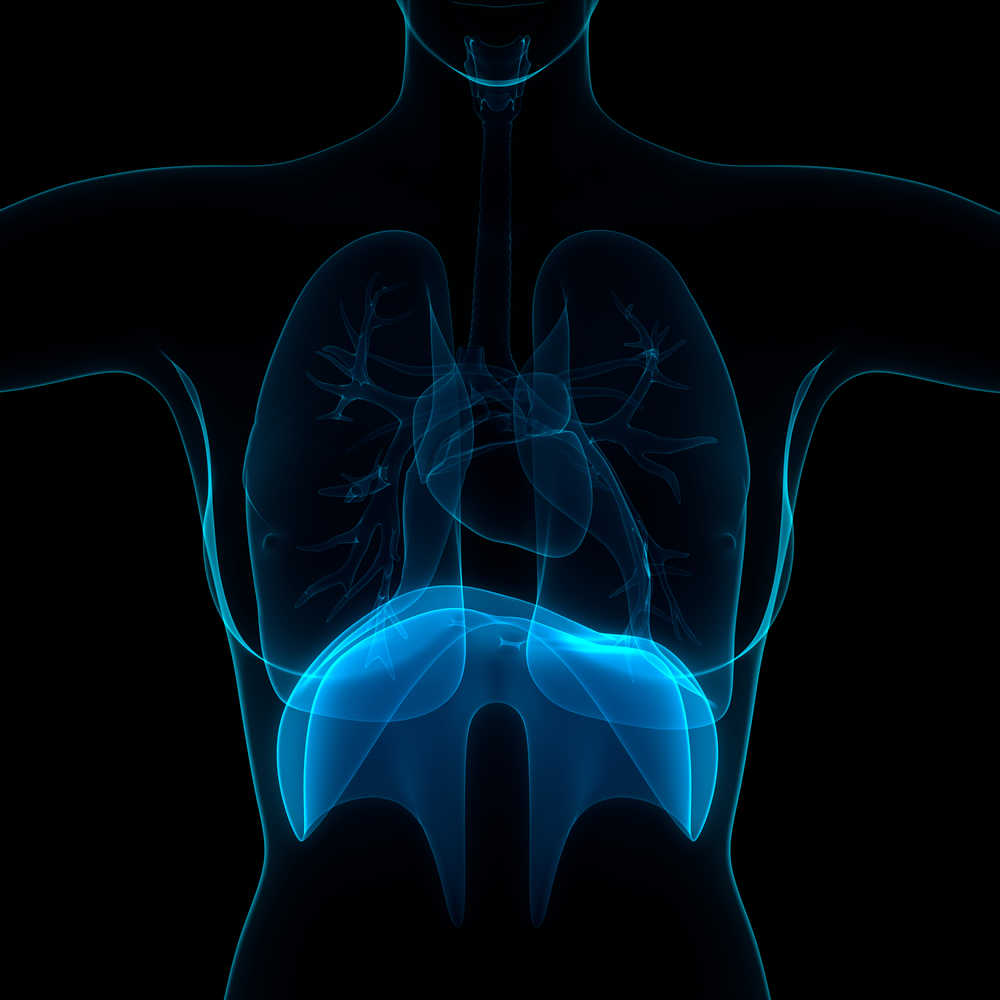
Diaphragm weakness may be assessed by nasogastric EMG or diaphragmatic ultrasound.
Diaphragm weakness may be due to ICU treatments:
• Sedation promotes disuse and may also have direct effects on the diaphragm
• Neuromuscular blockade renders the diaphragm inactive and causes atrophy
• Corticosteroids also contribute to muscle atrophy
• Mechanical ventilation further contributes to disuse atrophy and may cause load induced injury
Weakness is often also due to underlying disease processes:
• Hypercapnia and respiratory acidosis may reduce diaphragm contractility
• Cardiovascular shock may contribute via a mismatch of oxygen supply and delivery leading to anaerobic respiration and lactate production
• Sepsis causes altered distribution of blood flow, and cytokines such as TNF alpha directly impair contractile proteins
• Sepsis may also cause sarcolemma membrane fragility, increasing susceptibility to load related injury caused by mechanical ventilation
• Following surgery, diaphragm weakness is often due to stimulation of somatic or visceral phrenic afferent pathways or alternatively by direct nerve injury in cardiac surgery
Diaphragm weakness is associated with increased mortality and weaning failure. However weakness early in the ICU course may be due to underlying disease and it is unclear to what extent this predicts later weakness.
Maintaining spontaneous breathing is an important preventative measure, however this may be harmful in ARDS, although partial neuromuscular blockage and control of respiratory muscle activity may be possible.
Regarding treatment, hypophosphatemia should be corrected and evidence suggests theophylline or levosimendan may be effective. Inspiration training and diaphragmatic pacing are currently under investigation.
Take home messages
• Diagram weakness is common in intensive care
• It is associated with adverse outcomes including increased mortality
• The cause is often multi-factorial and contributed to by both underlying disease processes and ICU treatment
• Bedside techniques may aid diagnosis
• Maintaining spontaneous breathing during ventilation is the best evidenced preventative strategy, but partial control must be retained in ARDS
Article review was prepared and submitted by Matthew Harvey, Royal Brompton and Harefield NHS Foundation Trust on behalf of the EJRC.
Reference
Martin Dres, Ewan C. Goligher, Leo M. A. Heunks and Laurent J. Brochard. Critical illness-associated diaphragm weakness. Intensive Care Med (2017) 43:1441–1452, DOI 10.1007/s00134-017-4928-4